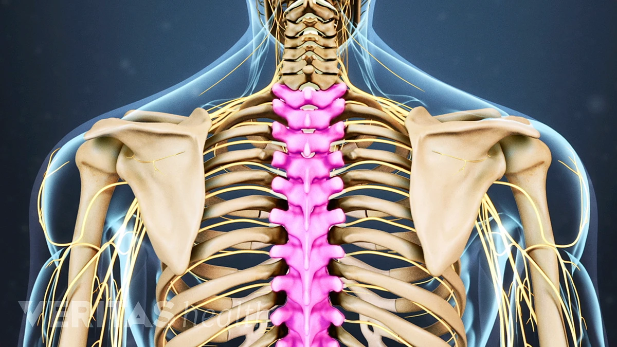
Back Side Body Structure | These roots exit the spine on both sides through spaces (neural foramina) between each vertebra. Kidneys filter waste out of the bloodstream, which is passed out of the body as. The ventral (from latin venter 'belly') surface refers to the front, or lower side, of an organism. At each level, a pair of nerve roots emerge from the right and left sides of the spinal cord. They are especially important to know if you plan to enter a healthcare field that involves analyzing images from mri machines and other types of imaging equipment.
The next pair of directional terms relate to the front or back of the body or structure, relative to another structure. It is the surface of the body opposite from the chest and the abdomen. On the same side of the body. The chest is inferior to the neck, and so on. Learn the lower back muscle anatomy associated with low back pain and hip pain.
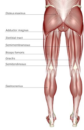
The cervical spine, the thoracic spine, and the lumbar spine. Learn the lower back muscle anatomy associated with low back pain and hip pain. The ventral cavity is on the front (anterior) of the body and is divided into the thoracic (chest) and abdominopelvic cavities. Part of the nerve that branches off the spinal cord or cauda equina. The muscles of the lower back, including the erector spinae and quadratus lumborum muscles, contract to extend and laterally bend the vertebral column. Human body anatomy female female anatomy muscle shoulder blade pain anatomy back muscles bones man female anatomy body muscles in a body female anatomy muscole shoulder concept muscular sysyem. Browse 385 human anatomy organs back view stock photos and images available, or start a new search to explore more stock photos and images. Involving both sides of the body. See human back anatomy stock video clips. The spine itself has three main segments: These muscles provide posture and stability to the body by holding the vertebral column erect and adjusting the position of the body to maintain balance. The vertebral column runs the length of the back and creates a central area of recession. The ventral (from latin venter 'belly') surface refers to the front, or lower side, of an organism.
Kidneys filter waste out of the bloodstream, which is passed out of the body as. All about the back muscles the back anatomy includes the latissimus dorsi, trapezius, erector spinae, rhomboid, and the teres major. If this vertical plane runs directly down the middle of the body, it is called the midsagittal or median plane. On this page, you'll learn about each of these muscles, their locations and functional anatomy. Most people have two kidneys, which are located near the back of the body, under the ribs, on each side of the spine.

These structures work together to support the body, enable a range of movements, and send messages from the. The ventral (from latin venter 'belly') surface refers to the front, or lower side, of an organism. The middle back, or thoracic spine curves outward. The eyes are superior to the mouth: Involving one side of the body. Download 208 human body outline front side back stock illustrations, vectors & clipart for free or amazingly low rates! These muscles provide posture and stability to the body by holding the vertebral column erect and adjusting the position of the body to maintain balance. Balance the weight of your head on top of your spine evenly distribute weights from your upper body into the lower extremities If talking about the skull, the dorsal side is the top. Roughly the size of a closed fist, each kidney measures about 10 to 12 centimeters long, 5 to 7 centimeters wide, and 3 to 5 centimeters thick. Scale to any size without loss of resolution. On the same side of the body. Your lower back (lumbar spine) is the anatomic region between your lowest rib and the upper part of the buttock.
The human back, also called the dorsum, is the large posterior area of the human body, rising from the top of the buttocks to the back of the neck. Summary the back consists of the spine, spinal cord, muscles, ligaments, and nerves. This curve, called lordosis, helps to: The ventral (from latin venter 'belly') surface refers to the front, or lower side, of an organism. Learn the lower back muscle anatomy associated with low back pain and hip pain.
New users enjoy 60% off. All about the back muscles the back anatomy includes the latissimus dorsi, trapezius, erector spinae, rhomboid, and the teres major. Browse 385 human anatomy organs back view stock photos and images available, or start a new search to explore more stock photos and images. These roots exit the spine on both sides through spaces (neural foramina) between each vertebra. The sagittal plane is the plane that divides the body or an organ vertically into right and left sides. Kidneys filter waste out of the bloodstream, which is passed out of the body as. On this page, you'll learn about each of these muscles, their locations and functional anatomy. Download this image now with a free trial. The muscles of the lower back, including the erector spinae and quadratus lumborum muscles, contract to extend and laterally bend the vertebral column. The chest is inferior to the neck, and so on. Download 208 human body outline front side back stock illustrations, vectors & clipart for free or amazingly low rates! In anatomy and physiology, the anatomical body planes and sections help us understand the various ways in which the body can be viewed when cut into sections. This curve, called lordosis, helps to:
Back Side Body Structure: Anatomically, the liver is divided into four unequal lobes.
Post a Comment